🔑 Key Learning
- Cardiac tamponade occurs when fluid builds up in the pericardial space, leading to diastolic compression of the heart and reduced cardiac output
- Presents with shortness of breath, tachycardia, and Beck’s triad: hypotension, raised JVP, muffled heart sounds
- ECG may show electrical alternans
- Treatment is urgent pericardiocentesis
🧬 Pathology
- Cardiac tamponade is caused by accumulation of fluid within the pericardial sac.
- As intrapericardial pressure rises above diastolic pressures, ventricular filling becomes impaired, resulting in reduced stroke volume and cardiac output.
- Common causes include trauma, myocardial infarction, malignancy, and post-cardiac surgery.

🩺 Clinical Features
- Symptoms: Shortness of breath
- Beck’s Triad:
- Hypotension
- Muffled heart sounds
- Raised JVP
- Additional findings:
- Pulsus paradoxus (drop in systolic BP >10 mmHg on inspiration)
- JVP with absent Y descent due to restricted right ventricular filling
🔬 Investigations
ECG may show:
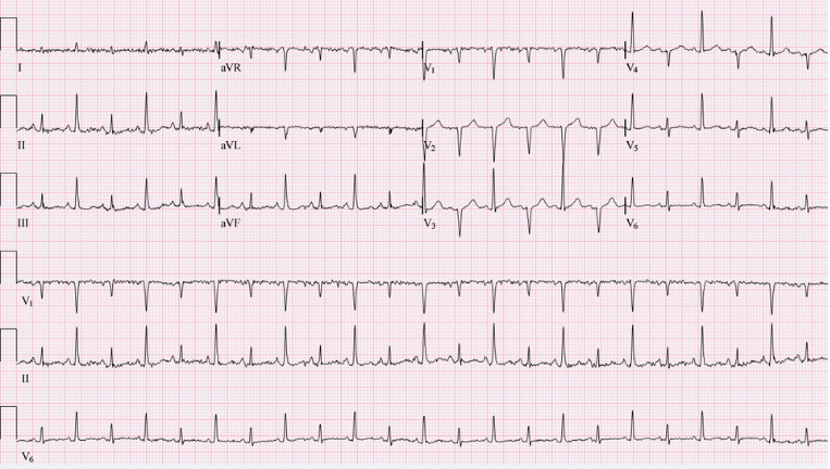
- Electrical alternans – alternating QRS amplitude beat-to-beat, due to swinging heart within the fluid-filled pericardium
💊 Management
- Urgent pericardiocentesis is required to relieve the pressure and restore cardiac output
📷 ECG Interpretation Practice
📝 Try to interpret the following ECG:
- What pattern do you see in the QRS complexes?