🔑 Key Learning
-
Dermatomyositis = Inflammatory myopathy + skin rash (photosensitive).
- Heliotrope rash, Gottron's papules
- Antibodies: Anti-Mi-2
-
Polymyositis = Inflammatory myopathy without skin involvement.
- Antibodies: Anti-Jo-1
- Dermatomyositis is a paraneoplastic syndrome in ~25%: associated with ovarian, breast, lung cancers. Consider screening for cancer.
🧬 Pathophysiology
- Autoimmune inflammation of striated muscle fibres ± skin.
- CD8+ T-cell-mediated muscle fibre damage in polymyositis.
- Complement-mediated microangiopathy in dermatomyositis, particularly affecting capillaries in skin and muscle.
👀 Clinical Features
Dermatomyositis
🧑🦰 Skin
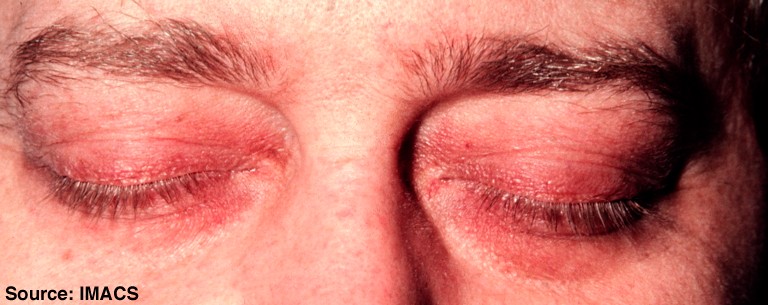
- Heliotrope rash: violaceous discolouration of eyelids
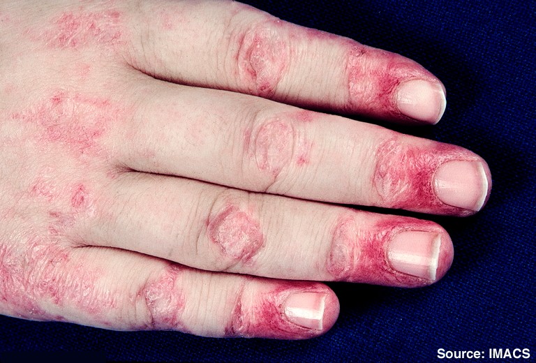
- Gottron’s papules: red/purple papules over knuckles, PIP, DIP
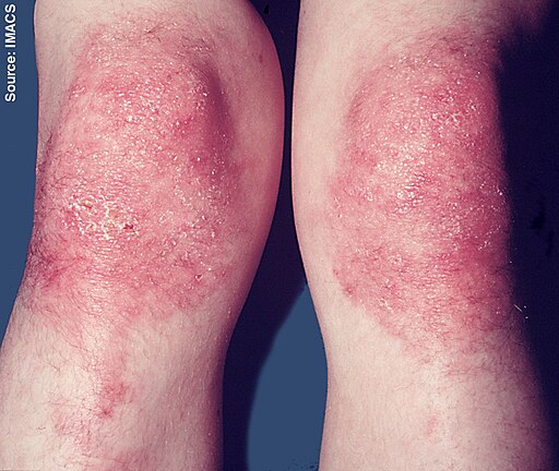
- Gottron’s sign: erythematous rash over extensor surfaces (e.g. knees, elbows)
- Photosensitive red macular rash over shoulders and back (‘shawl sign’)
- Mechanic’s hands: dry, cracked skin on hands
💪 Muscle
-
Symmetrical proximal muscle weakness
- Exam MCQs may describe difficulty climbing stairs, rising from chair, lifting objects, brushing hair
⚠️ Systemic
- Weight loss, fatigue, night sweats
- ILD: dry cough, SOB
- Raynaud’s phenomenon
- Malignancy risk: screen for ovarian, breast, lung, GI cancers


Polymyositis
- Similar proximal muscle weakness as dermatomyositis, but no skin features
- Can occur in association with connective tissue diseases (SLE, systemic sclerosis)
🧪 Investigations
Bloods
- Raised CK and other muscle enzymes (LDH, ALT/AST)
- Consider muscle biopsy
- ANA positive (~80%)
-
Autoantibodies:
- Anti-Mi-2 → Dermatomyositis
- Anti-Jo-1 → Polymyositis
💊 Management
- 1st Line: High-dose corticosteroids (e.g. prednisolone)
- Immunosuppressants: Methotrexate or azathioprine if inadequate response
- Screen for malignancy in dermatomyositis (especially if age > 40 or rapid onset)
📝 Exam Clues & Clinchers
- Heliotrope rash + proximal weakness + Shawl sign = dermatomyositis
- Elevated CK + anti-Jo-1 + ILD = polymyositis
- Associated cancers: ovarian, breast, lung, GI
- Gottron’s papules = red, scaly bumps over knuckles
- Anti-Mi-2 = Dermatomyositis
- Anti-Jo-1 = Polymyositis
